Paramecium.jpg
sur le Dico des Ados : ton dictionnaire collaboratif, libre et gratuit !

Taille de cet aperçu : 610 × 599 pixels. Autres résolutions : 244 × 240 pixels | 488 × 480 pixels | 751 × 738 pixels.
Fichier d’origine (751 × 738 pixels, taille du fichier : 186 kio, type MIME : image/jpeg)
Description
| Description |
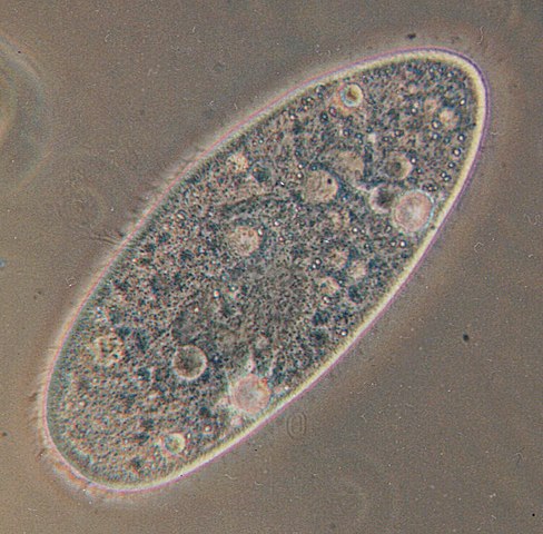
Deutsch: Paramecium aurelia - Optisches Mikroskop. Paramecium aurelia, der bekannteste von allen ciliaten. Die Blasen innerhalb der Zelle sind Vakuolen. Die gesamte Oberfläche ist mit Wimpern umgeben, die durch ihre schnelle Bewegung verwischt werden.
English: Paramecium aurelia. Optical microscope. Paramecium aurelia, the best known of all ciliates. The bubbles throughout the cell are vacuoles. The entire surface is covered in cilia, which are blurred by their rapid movement.
Français : Paramecium aurelia. Microscope optique. Le plus connu des ciliés. Les bulles que vous voyez sont des vacuoles. Tout le corps est couvert par des cils, qui sont flous sur l'image à cause de leurs mouvements rapides.
Polski: Paramecium aurelia - pantofelek, najbardziej znany ze wszystkich orzęsków. Bąbelki w środku komórki to wodniczki. Cała powierzchnia pantofelka pokryta jest rzęskami, które są na fotografii zamazane ze względu na ich szybki ruch.
Српски / srpski: Paramecium aurelia, najpoznatiji od svih trepljara pod optičkim mikroskopom. "Mehurići" u ćeliji paramecijuma su vakuole. Cela površina tela je prekrivena trepljama, koje su na slici mutne zbog toga što se brzo pokreću.
Türkçe: Paramecium aurelia - optik mikroskop. Paramecium aurelia, tüm siliyalılar içinde en çok bilinen türdür. Hücre boyunca yuvarlak olarak izlenen oluşumlar, vakuollerdir. Hücrenin tüm yüzeyi, hızlı hareketlerinden dolayı bulanık görüntü vermiş olan siliya ile kaplıdır. |
| Date | |
| Source | Originally uploaded to the English Wikipedia, where it was made by Barfooz. |
| Auteur | Barfooz at the English Wikipedia. |
| Autres versions | Transparent |
Conditions d’utilisation

|
Vous avez la permission de copier, distribuer et modifier ce document selon les termes de la GNU Free Documentation License version 1.2 ou toute version ultérieure publiée par la Free Software Foundation, sans sections inaltérables, sans texte de première page de couverture et sans texte de dernière page de couverture. Un exemplaire de la licence est inclus dans la section intitulée GNU Free Documentation License. |
| Ce fichier est disponible selon les termes de la licence Creative Commons Attribution – Partage dans les Mêmes Conditions 3.0 (non transposée). | ||
| ||
| Ce bandeau de licence a été ajouté à ce fichier dans le cadre de la procédure de mise à jour des licences des images sous GFDL. |
Soft scrubbed view
Journal des téléversements d’origine
Originally uploaded to English Wikipedia.
- 23:11, 27 October 2004 . . Barfooz (Talk) . . 751x738 (190517 bytes) (Paramecium viewed under a microscope)
- 15:19, 28 June 2004 . . Josh Grosse (Talk) . . 236x152 (3913 bytes) (Reverted to earlier revision)
- 15:19, 28 June 2004 . . Josh Grosse (Talk) . . 236x152 (5129 bytes) (Reverted to earlier revision)
- 15:13, 28 June 2004 . . Josh Grosse (Talk) . . 236x152 (3913 bytes) (Better image, created by self)
- 20:04, 10 October 2003 . . Josh Grosse (Talk) . . 236x152 (5129 bytes)
Historique du fichier
Cliquer sur une date et heure pour voir le fichier tel qu'il était à ce moment-là.
| Date et heure | Dimensions | Utilisateur | Commentaire | |
|---|---|---|---|---|
| actuel | 31 mai 2005 à 22:46 | 751 × 738 (186 kio) | wikimediacommons>Luis Fernández García | ''Paramecium aurelia''. Optical microscope Source: English Wikipedia (http://en.wikipedia.org/wiki/Image:Paramecium.jpg) |
Utilisation du fichier
La page suivante utilise ce fichier :
La liste (mise en cache) risque d’être non actualisée. Pour voir la dernière version, connecte-toi.